
Gillette Children's
Hospital and Clinics
For more than 125 years, we’ve focused on some of the toughest challenges in pediatric medicine. We care for brain, bone and movement conditions needing specialized expertise. When families arrive at our door, they feel they’ve come home. We’re inspired by their perseverance.
Developmental Evaluations
For infants and toddlers at higher risk of developmental delays.
Understand Better
gilletteSTORIESonline.
-
 April 25, 2024
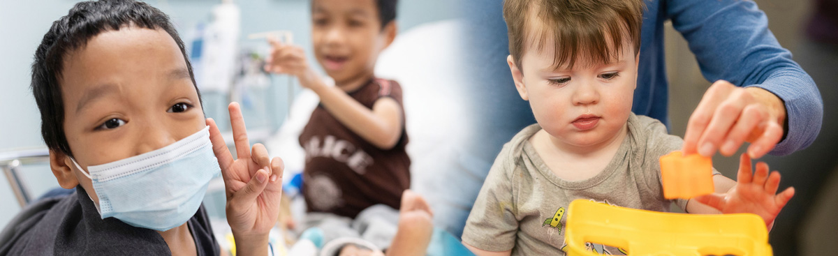
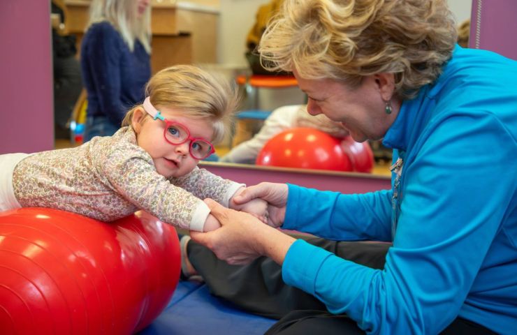
Occupational Therapists (OTs) Help Infants and Toddlers Thrive
Parents concerned about a child exhibiting differences in movement, play, learning, or communication can get…
-
 April 22, 2024
Gillette Children’s Champions Environmental Sustainability
Gillette’s commitment to environmental sustainability is deeply rooted in our work, with a focus on improving…
-
 April 15, 2024
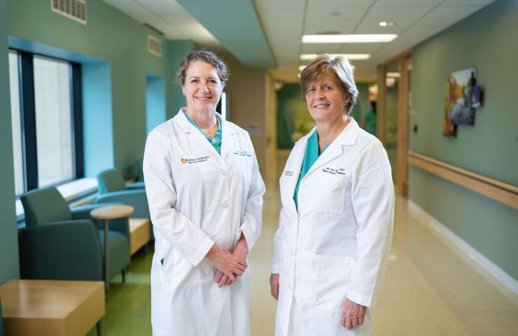
Meet the Physicians Creating Better Understanding of Limb Differences
April marks Limb Loss and Limb Difference Awareness Month, a time dedicated to raising awareness…
-
 April 11, 2024
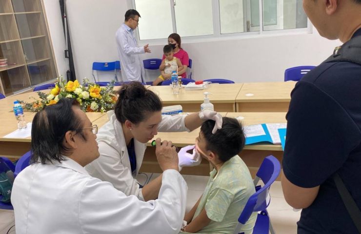
Bringing Smiles and Surgical Skills to Vietnam
Three Gillette Children’s surgeons recently traveled to Vietnam on a mission trip to help physicians in…
-
 March 21, 2024
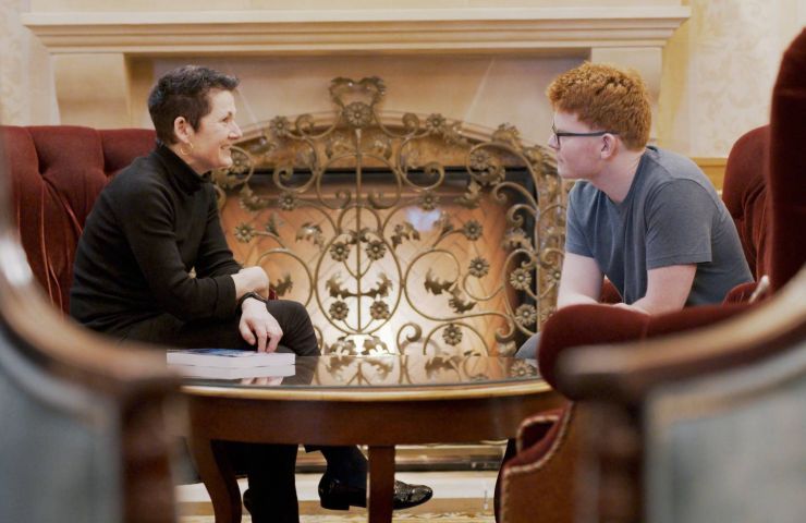
How do you beat your disability? By never letting it define you.
Featured in the book by Lily Collison (in collaboration with experts at Gillette) is her…
-
 March 19, 2024
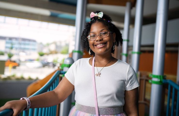
Gillette Children’s Announces 2024 Children’s Miracle Network Champion
ZaLayaa Jahzara Wandrick is 11 years old. She’s an excellent cook, crafter and loves science, soccer…