Orthopedics Researchers Benefit from Multimillion-dollar Grant
Gillette Children’s and the University of Minnesota have a rich research collaboration history that continues to expand with the arrival of a new grant from the National Institutes of Health (NIH).
With a shared interest in studying developmental joint disorders, a team of experts from Gillette and the University have established a new collaboration to study Legg-Calvé-Perthes disease. This new project was developed under the leadership of Jennifer Laine, MD, Medical Director of Research at Gillette; Susan Novotny, PhD, Clinical Scientist at Gillette; and Casey Johnson, PhD, Assistant Professor of Medical Imaging at the University of Minnesota. Dr. Johnson, a magnetic resonance imaging (MRI) research scientist, was awarded a $2.7 million NIH R01 grant to investigate non-invasive imaging methods for early detection and monitoring of osteonecrosis of the femoral head, with a particular focus on Legg Calvé-Perthes disease.
The collaborative research project will address a critical need for better imaging to inform treatments for early stage Perthes. Dr. Laine says: “The hope is that this grant will allow us to develop advanced techniques that are sensitive enough to assess bone ischemia, necrosis, and repair. With these advances, we anticipate better understanding of the underlying disease and the way the hip heals. This will help inform treatment decisions and assess response to treatment.”
Dr. Johnson is excited at the prospect of developing new imaging approaches. He says: “Our proposal is a critical step toward improving long-term outcomes for children and young adults suffering from osteonecrosis of the femoral head. With this study, we will advance new imaging solutions that allow for detection and monitoring of Perthes disease in its earliest stages, when it is most amenable to treatment.”
Osteonecrosis and Perthes Disease Care at Gillette
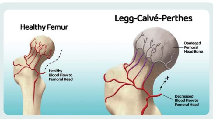
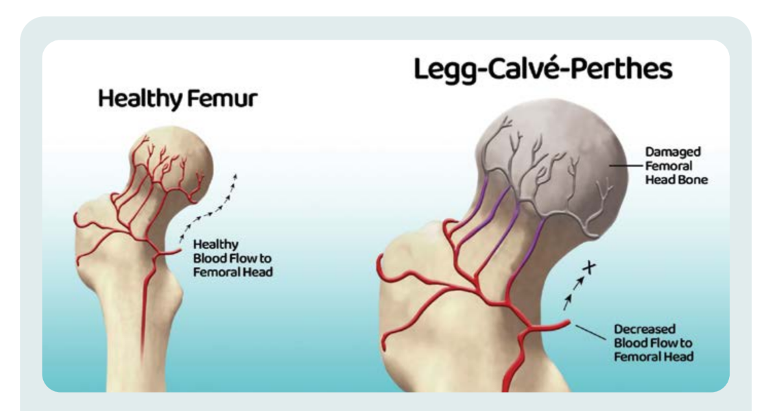
Perthes disease, a type of osteonecrosis of the femoral head, is a painful hip disorder that severely impacts the lives of children and young adults, and can lead to lifelong hip dysfunction and chronic pain. Perthes is caused by an interruption of blood supply to the hip, which causes death of the bone and cartilage in the hip.
Over time, the body absorbs the dead bone and builds new bone. As the hip goes through these processes, it can become fragile and at risk for damage, sometimes losing the spherical ball and socket shape of the hip joint. Currently, Perthes treatment focuses on maintaining the spherical shape of the hip joint to mitigate the condition. Despite significant research, many big questions remain regarding Perthes disease, including why it happens, how to facilitate the healing process, and how to treat it most effectively. Perthes disease, therefore, is one of the most difficult and controversial conditions treated in pediatric orthopedics. This is why researchers are dedicated to finding better answers for patients and families impacted by this condition.
Join Our Partners in Care Community!
Subscribe to Partners in Care Journal, a newsletter for medical professionals.
Subscribe Today